Eye Worms
These worms live in the conjunctival sac (eyelid) of the eye in many species of livestock, in many countries. Cattle, sheep, horses, camels, goats, pigs, dogs, cats, birds, and humans can be affected. The most common species in Kenya is Thelazia rhodesii, which parasitizes cattle and sheep. The worms are up to 2cm long and are thin and white. One or both eyes may be affected.
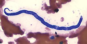 |
| Eye Worm |
|
© Courtesy of the United States Federal Government
|
Mode of Spread
Various species of fly, in which the worm has part of its life cycle, are responsible for the spread of the worm from one animal to another. The fly has a preference for eye secretions which are ideal for transmission. The fly ingests the larvae which become infective in 2-4 weeks. These larvae are mechanically deposited in teh host's eye by the fly during feeding. Development of sexually mature worms takes about 1-4 weeks in cattle.
The worm lives under the eyelids, in the conjunctival sac and under the third eyelid. The worm has a rough cuticle (skin) which causes irritation and inflammation to the cornea.
Signs of Eye Worm
- Excessive production of tears which may contain pus.
- Avoiding sunlight.
- Inflammation of the thin membrane covering the white of teh eye and the inner surface of the eyelid (Conjunctivitis).
- Cloudiness of the cornea and sometimes ulceration and piercing of the cornea.
- Lack of response to treatment with antibiotics.
Diagnosis
Close inspection of the eyes will reveal small white worms swimming in the conjunctival fluid. Several animals should be examined as it will not be possible to see worms in every animal. Even if the worms can be seen with the naked eye, a veterinarian should be consulted to confirm their presence.
Prevention of Eye Worms Infestation
The condition is less severe than Pink Eye. Control of the fly is not practical.
Recommended Treatment and Control
- A qualified veterinarian can remove the worms with forceps after instillation of a local anaesthetic solution or
- Using an eye wash containing a local anaesthetic and wash the worms out of the eye. A mixture of 10 ml of 2% local anaesthetic solution with 40-50ml of clean water makes the eye wash. 5-10 ml is put onto the eye and after waiting for about 2 minutes, the worms are washed out of the eye using clean cold water.
- Certain systemic dewormers such as Levamisole at 5 mg/kg under the skin and Ivermectin and Doramectin both at 0.2 mg/kg SC or in the muscle are effective treatments.
- Pour-on forumlations of Ivermectin and Doramectin, delivered on the back of the animal to achieve a dosage of 0.5 mg/kg are also highly effective.
- Treatment is also possible with topical application of Levamisole or topical Ivermectin. Bother are given as a 1% aqueous solution directly into the eye. If the eye discharge is cloudy or white it may be advisable to put an antibiotic ointment into the eye following removal of the worm or following the administration of dewormers.