East Coast Fever Complex (Theileria parva parva, Theileria parva lawrencii)
Local names:
Gikuyu: ngai / Kamba: ngai / Embu: ngai / Gabbra: shilmi dimtu / Maasai: oltikana, oldigana / Kipsigis: sosoito / Swahili: ndigana / Turkana: lokit, loleeo, lipis / Iteso: angaarwei / Luidakho: yapwolo / Luvugusu: gamavumba / Nandi: cheptigonit / Pokot: cheptikon, lipis / Maragoli: ivitu, evivitu / Meru: ita / Samburu: lipis / Borana: lipis / Somalia: udhurshillin kener
Common names:
Theileriosis, African coast fever, la fievre de la cote orientale (French), fiebre de la costa oriental Africana (Spanish)
Description:
Tick borne protozoal parasite disease
Introduction
East Coat Fever is a non-contagious fever-producing disease of cattle transmitted by the brown ear tick.
"Classical" East Coast Fever is caused by a protozoal parasite closely related to Babesia called Theileria parva parva, which invades the lymph nodes. It is transmitted by brown ear ticks (Rhipicephalus appendiculatus) and characterised by high fever, an abnormal lowering of the white blood cell count and severe damage to the lymphoid system. The disease is localised to East and Central Africa - Kenya, Uganda, Tanzania, Rwanda, Burundi and Malawi with extensions into the Congo, Zambia, Southern Sudan and Mozambique.
Corridor Disease is caused by Theileria parva lawrencii and is transmitted from buffalo to cattle by Rhipicephalus appendiculatus in Eastern Africa and also by Rhipicephalus zambezienis in the low veldt of Central Africa. It is called "Corridor" Disease as it was first discovered in a corridor of land between the Umfolozi and Hluhluwee Game Reserves in Natal in South Africa. When the disease appears in areas where there is chance of close contact between buffalo and cattle, usually at forest edges, on uncleared land or in areas of dense bush, cattle keepers should expect occurrence of corridor disease.
Other less infectious forms of the disease are widespread in tropical and subtropical regions of Africa, Asia and southern Europe. These include T.parva bovis, mutans and taurotragi.
Mode of spread
"Classic" East Coast Fever is transmitted from cattle to cattle by the Brown Ear Tick, Rhipicepahalus appendiculatus. The distribution of East Coast Fever is geographically confined by the distribution of the Brown Ear Tick and by whether the tick can undergo more than one life-cycle per year. This confines it to areas of rainfall of more than 750mm per year and where it is not too cold or high. There are large areas of Africa where the tick is present but not the disease. There are no areas where the disease is present but not the tick.
Corridor Disease is not normally maintained in cattle. Close contact is required between buffalo and cattle, usually at forest edges, on uncleared land or in areas of dense bush.
Incubation varies between 9 to 18 days, most commonly 14 to 16 days after exposure to infective ticks.
Parasites in the blood of an infected animal are picked up by immature stages of the tick. These complete feeding, drop off and moult and then, at the next stage in their life stage, attempt to feed. If picked up by a susceptible animal, the parasites in their salivary glands complete their development over the 3 days after attaching and, between the third and fifth day of feeding, the tick secretes infective sporozoites into the new host.
One tick will transmit sufficient sporozoites to kill a susceptible cow. Both European and local breeds from areas where the disease is not endemic are susceptible but Zebu cattle are more resistant. This, coupled with their relative resistance to ticks, makes them significantly more resistant to East Coast Fever than European breeds. However, Zebu cattle from areas where there is no East Coast Fever are equally susceptible to infection.
Infected ticks may live for 1 - 2 years but normally they lose the infection in about 11 months. Infected and clean ticks do not move, although they may be carried by animals or man. If cases occur on a farm it must be remembered that further cases may occur during the next year. A close watch must be kept at all times. There is no room for relaxation of standards.
Clinically infected cattle, exhibiting the parasites, are the chief source of infection for the tick. In classical East Coast Fever cattle do not remain as carriers but exceptions are now reported with increasing frequency and effective disease control programs have been based on this premise.
In East Coast Fever enzootic (prevalent) areas a high proportion of cattle naturally acquire immunity to the disease when first exposed as young calves of immune dams. This immunity effectively protects them against an otherwise lethal challenge. This, however, will not occur in non-enzootic (non-prevalent) areas.
Signs of East Coast Fever
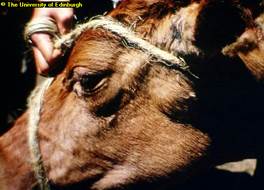
- The parotid lymph nodes below the ear become enlarged 14 to 16 days after attachment of the tick.
- A few days later a fever develops. This can be up to 41 degrees Celsius to 42 degrees Celsius (106 degrees Fahrenheit to 107 degrees Fahrenheit).
- Milk yield decreases
- Other superficial lymph nodes enlarge, especially those in front of the shoulder joint and in front of the stifle joint of the hind leg. These are often clearly visible to the attentive observer.
- Appetite decreases, body condition declines, parasaties appear in the blood and the animal is infective to ticks and hence to other animals
- There is fluid in the lungs and pneumonia occurs. The animal breathes increasingly rapidly and with increasing distress. It starts to cough, especially if made to move and nasal discharge is common.
- The eyes often appear milky or blue and there may be shedding of tears.
- Sometimes there is diarrhoea which can be violent or blood stained.
- There is muscle wasting and posterior weakness
- Finally the animal collapses and dies, usually about 18 to 26 days after tick attachment.
 |
 |
|
Swollen Lymph node as sign of ECF carried by the brown ear tick shown below |
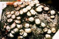
Brown ear ticks on the ear of a calf |
| © The University of Edinburg |
© The University of Edinburg
|
Diagnosis
Very often when an animal has died from East Coast Fever or Corridor Disease there is a pile of white froth lying on the ground in front of its nostrils. The sight of this should always stir an observant person into thinking about ECF/Corridor Disease. This froth is due to the massive pulmonary oedema (lung fluid) which has killed the animal.
Post mortem examination will reveal lungs full of fluid and the windpipe full of white froth. Cutting the lungs with a knife will release a flood of fluid from the now clearly defined pulmonary lobules. This is a dramatic, almost diagnostic sign.
Lymph nodes are enlarged and fluid-filled. The kidneys may have white spots on their surface - "infarcts". In the abomasum or fourth stomach may be seen punched out erosions.
A final diagnosis is dependent on identifying the parasite in the animal. Blood smear and gland (biopsy) smears should be taken for laboratory analysis by a qualified vet to confirm the disease.
The overall picture should be taken into consideration. A change in some aspect of management some 2 to 3 weeks earlier, a change of acaricide (tick-killing solution), the appearance of ticks, increased rainfall or improved ground cover leading to extended tick survival, the intrusion of buffalo, illegal cattle movement within the area, all must be considered when formulating a strategy for control.
Apart from the clinical picture, post mortem findings and the examination of blood and lymph node smears there are a number of serological diagnostic tests, but these are of limited value in the field.
Diseases with similar symptoms
Enlarged lymph nodes, coughing, watery eyes and discharge from the nose: See Trypanasomiasis and Malignant Catarrhal Fever.
Prevention - Treatment - Control
Prevention/Control
- The infection and treatment method of immunisation against East Coast Fever is gaining ground. The antibiotic used is a long acting (LA) Oxytetracycline, which inhibits development of the parasite when given at the onset of infection. The animal is injected with live vaccines of East Coast Fever. This will confer protection to this strain for more than 3 years. However there are different strains of East Coast Fever and when immunised cattle are moved to a different area they may not be protected. Likewise if infected cattle from another area move in to a property they may bring in with them other strains of East Coast Fever.
- Effective Tick control. Tick control continues to be the most important measure in the fight to prevent cattle from becoming infected with East Coast Fever. The Brown Ear Tick is a three host tick i.e. it does not spend its entire life on the animal but drops off between the various stages of its life cycle; therefore when an outbreak occurs, either on the property or in the immediate area, once weekly dipping or spraying will not be sufficient. Dipping or spraying must be done twice weekly to break the transmission cycle between cattle and tick and continued as long as is necessary, for example if the tick problem continues to be unacceptably high. Pyrethrum grease should be applied to the inside of the ears and below the base of the tail, whose end should be clipped. All of this is costly, but not as costly as losing valuable stock or having to use very expensive curative drugs.
- Newer tick control measures include the Spot-on / Pour-on drugs
Treatment
Treatment of East Coast Fever / Corridor Disease is effective but it MUST be started EARLY in the disease process by a qualified veterinarian. Once the disease is well established treatment may not be effective. Available curative drugs are very expensive so every effort must be made to prevent infection from becoming established in the first place.
The drugs available for treatment are Parvaquone ("Clexon"), Buparvaquone ("Butalex" and others) and Halofuginone ("Terit"). The first two are given by intramuscular injection, the last by mouth. The dose for Paravaquone is 20 mg/kg bodyweight, Buparvaquone 5mg/kg bodyweight and Halofuginone 1 mg/kg bodyweight. If there is no satisfactory response to treatment after 48 hours this treatment should be repeated. Together with one of these drugs the affected animal should be given Oxytetracycline by intramuscular injection, preferably a long-acting formulation to lessen the number of times the animal is handled and to lessen the number of injections given. A dose of 30 mg/kg bodyweight should be given and under-dosage should be avoided. Most people tend to underestimate body weights. This must be avoided, both to ensure that the animal is given the correct dose and to avoid causing drug resistance.