Pink Eye
Pink Eye or Infectious Kerato conjunctivitis is an infection of the eyes of cattle, sheep, and goats with a mixture of micro-organisms which include Moraxella Bovis, Mycoplasma species, Listeria monocytogenes and Chlamydia.
The infection occurs in animals of all ages but is more severe in young animals. The disease is generally less severe in sheep and goats. One or both eyes may be affected. Zebu cattle appear to be less likely to be affected than European breeds of cattle.
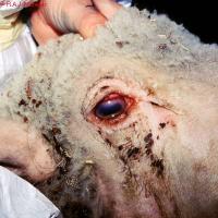 |
| Keratoconjunctivitis in a Sheep caused by MmmLC |
|
© R.A.J. Nicholas. Reproduced from the Animal Health and Production Compendium, 2007 Edition. CAB International, Wallingford, UK.
|
Mode of Spread
- Moraxella bovis is the organism primarily responsible for the severest form of the disease in cattle.
- When cattle are eating silage, Listeria may cause conjunctivitis, but less severe in than when caused by Moraxella.
- In sheep Clamydia is the most common cause. The disease is generally less severe in sheep and goats.
- Infection does not spread between small ruminants (like sheep and goats) and cattle since the causative agents of the disease for small ruminants differ from those which affect cattle.
- Spread by direct contact with infected animals, especially when animals are crowded together.
- In cattle, dry dusty conditions, shipping stress, irritants such as grasses and pollens, grasses contaminated by eye discharges, bright sunlight, lack of pigment in the eyelids and flies may increase disease occurrence.
- Flies also spread the disease in cattle.
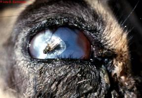 |
| Pink Eye is spread by flies |
|
© John B. Bashiruddin. Reproduced from the Animal Health and Production Compendium, 2007 Edition. CAB International, Wallingford, UK.
|
Signs of Pink Eye
- The incubation period is usually 2-3 days but can be up to 3 weeks after infection.
- One or both eyes may be affected.
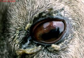
- The eye becomes watery, the lining under the eyelid becomes red and inflamed and the animal blinks repeatedly and diverts the affected eye(s) away from bright sunlight.
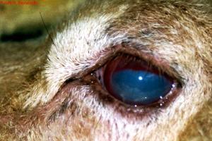
- A small unclear area appears in the centre of the eye. Within a few hours a faint haye appears which becomes denser. After 48-72 hours the whole cornea may be unclear, blinding the animal in that eye. If the eye is hazy white to blue this is due to accumulation of fluid. If it is milky white or yellow this is due to white blood cell infiltration and indicates severe infection.
 |
 |
|
| Pink eye signs - a small unclear area in centre of the eye | Pink Eye signs | |
|
© John B. Bashiruddin. Reproduced from the Animal Health and Production Compendium, 2007 Edition. CAB International, Wallingford.
|
© John B. Bashiruddin. Reproduced from the Animal Health and Production Compendium, 2007 Edition. CAB International, Wallingford. |
 |
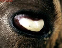
| Thickened corenea after healing of Pink Eye. |
|
© John B. Bashiruddin. Reproduced from the Animal Health and Production Compendium, 2007 Edition. CAB International, Wallingford.
|
 |
|
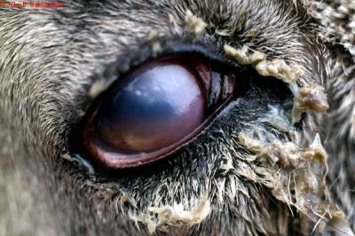
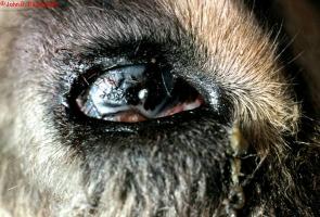
| Severe Pink Eye | |
|
© John B. Bashiruddin. Reproduced from the Animal Health and Production Compendium, 2007 Edition. CAB International, Wallingford.
|
 |
| Ruptured cornea after pink eye |
|
© John B. Bashiruddin. Reproduced from the Animal Health and Production Compendium, 2007 Edition. CAB International, Wallingford. |
- Blood vessels invade from the edge of the cornea.
- A small ulcer is apparent near the centre of the cornea.
- Continued active ulceration may cause the cornea to rupture.
- Appetite is often suppressed due to pain and visual impairment, milk yield is likely to drop and there may be loss of weight.
- Most animals slowly recover over a period of about a month, sometimes a small white scar remains. About 2% are left blind in the affected eye. In severe cases the eye becomes conical in shape, with ulceration at the tip. These eyes are liable to rupture.
Other conditions can cause inflammation of the eye, so in every case, a close inspection of the affected eye is essential.
- Foreign bodies, such as grass seeds and awns can become stuck to the surface of the eye.
- Thorns can be embedded in the cornea.
- Spitting cobras can project venom into the eye.
- Corrosive fluids and saps can inflame the cornea.
- Worms can live in the conjunctival sac.
Prevention and Control of Pink Eye
- Avoid overcrowding of animals.
- Sick animals should be isolated and treated.
- Affected animals should have access to shade.
- Cleanliness in the cattle yards should be maintained in an effort to keep fly numbers to a minimum.
- Insecticides in the form of pour-ons may be beneficial.
- Recovered animals have an immunity lasting for about 12 months.
Recommended Treatment
Although many cases do recover spontaneously this is a potentially serious condition and therefore treatement should be considered before ulceration of the cornea leads to permanent blindness. A variety of antibiotics can be prescribed by a qualified veterinarian. These can be given by injection or in the form of eye ointment.
- Oxytetracycline is the drug of choice for systemic treatment as it is concentrated in corneal tissue. Two injections (20mg/kgIM) of a long-acting oxytetracyline forumlation (-200mg/ml) at 72 hour intervals is the treatment of choice (as per the manufacturer's instructions).
- Ampicillin, penicillin, gentamycin and kanamycin can be injected into the eye, sometimes in conjunction with a corticosteroid such as dexamethasone, but this should be done by a skilled veterinarian.
- Eye ointment require frequent application to be effective, as often as three times daily. Oxytetracycline/polymixin B, gentamycin and erythromycin are effective. The ointment must be instilled carefully under both upper and lower eyelids.
- In severe cases the use of 1% atropine drops or ointment will prevent body spasms within the eye and reduce the likelihood of stickiness forming within the eye.
- If an ulcer ruptures the eye will collapse and shrink. In some cases there is continuing discomfort, pain and discharge due to ongoing infection. In many cases removal of the eye by a vet is the best option. This involves removal of the eye, leaving the eye muscles and remaining orbital contents intact.