This datasheet guides to diseases judging from symptoms in animals, it is a tool to assist farmers find out what may be wrong when their animals look unwell or die suddenly. It aso provides guidance on how to submit samples from sick animals (or whole dead animals) to a laboratory for analysis.
It further contains the Diagnostic chart for Cattle and the Diagnostic chart on Sheep & Goats wich guides the identification of diseases
Introduction - signs of disease
Farmers and pastoralists know that animals are sick when they notice changes in behavior such as refusal to eat, keeping to shady areas, or physical signs such as different breathing, coughing, body swellings and weakness etc.
Serious livestock farmers will keep observing their animals on daily basis to make sure no such signs miss their attention. It is important to catch such symptoms at an early stage in order to treat before the disease becomes too serious to treat.
The below guide to diseases judging from symptoms is a tool to assist farmers find out what may be wrong when their animals look unwell or die suddenly. It aso provides guidance on how to submit samples from sick animals (or whole dead animals) to a laboratory for analysis.
Animals cannot speak and tell us where they hurt. But we can observe the vital functions of their body and their behaviour. Feeding and ruminating are the best indicators of good health. For the good observer slight changes in feeding and ruminating can indicate beginning of a disease and early action can be taken (e.g. measuring the temperature). Th earlier you are aware that an animal is sick the earlier you can start treatment and the more successful your treatment is going to be. – Treating animals that have been sick for long (chronic cases) is very difficult, costly and often a complete waste of time.
Disease Signs include:
- Loss of appetite or not feeding at all
- Fever
- Abnormal consistent of the faeces
- Abnormal colour (or consistence) of the urine
- Abnormal colour or consistence of the milk
- Swollen and hot areas of the body such as lymph glands or the udder
- Breathing rate
- Unusual smells
- Abnormal behaviour
1. Body temperature and fever
Normal body temperature varies by about 0.5°Celsius during the day and can be a bit lower (early morning) or a little bit higher (evening) than the normal body temperatures listed in the following table. To measure body temperature you need a veterinary thermometer. It is very cheap and can be found in most agro-vet shops. It is an essential tool for theserious livestock farmer.
Body temperature in animals
|
Type of Animal |
Normal Body temperature in °C |
Upper limit in °C (any higher temperature is fever*) |
|
|
|
|
|
Cattle |
38.5 |
39.5 |
|
Calves |
39.0 |
40.0 |
|
Horses, mules, donkeys |
38.0 |
39.0 |
|
Foals |
38.5 |
39.5 |
|
Sheep |
39.0 |
40.0 |
|
Goats |
39.5 |
40.5 |
|
Pigs |
39.0 |
40.0 |
|
Piglets |
39.5 |
40.5 |
|
Rabbits |
39.0 |
|
|
Dogs |
38.5 |
|
|
Cats |
38-39 | 39.5 |
|
Birds |
40.5 |
|
Adapted from Blood Radostits Henderson
2. Breathing rate , lung noises and other internal sounds
 |
| Stetoscope |
|
(c) William Ayako, KARI Naivasha
|
 |
| Use of a Stetoscope |
|
(c) William Ayako, KARI Naivasha
|
Normal breathing rates for animals
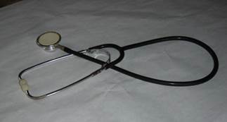
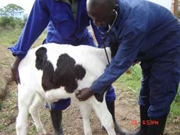
You can count the breathing rate of a sick animal by standing next to it and counting the breathing movements for one or two minutes (inhaling + exhaling together count as one breath). There are different sounds generated inside the body of animals (like heart beat, breathing, stomach sounds), which are not easy to hear. Veterinarians (and human doctors) use a stethoscope that magnifies these sounds. Placing your ear on the skin of the chest wall of the animal above the lungs can help hearing abnormal lung noises in case of pneumonia. Veterinarians are trained to listen for abnormal sounds, so if there is a problem it is usually best to call the vet. However, keen farmers can also buy a stethoscope from an agro- vet shop and try to learn to use it.
Breaths per minute of healthy adult animals
|
Healthy adult |
Breaths per minute |
|
Cattle |
12 |
|
Sheep,goats |
12 |
|
Horses |
12 |
|
Mules,donkeys |
12 |
|
Camels |
10 |
|
Pigs |
15 |
|
Cats |
20-30 |
|
Dogs |
20 |
3. Observing and Describing Disease Signs
The following list of symptoms intends to help you in recognizing disease and to also describe the disease signs to others (e.g. to the vet over the phone):
Fever: measured by thermometer in degrees Celsius(shivering is often a sign of fever)
Breathing: dilated nostrils, facing the wind, groaning, grunting, coughing
Face Expression: off feed, nervous, excitable, aggressive, dull, lethargic
Nose/Nostrils: dry nose, running nose: watery fluid, pus, bloody fluid
Body condition: weak, thin, emaciated
Skin: matted color, dry, rough, peeling, scruffy, crusted, lumpy, bald, lesions and swellings on the skin surface, bearing lice/ticks/fleas
Mucous membranes: (these are white skin areas inside the eyelids below the eyeball, and the inside of the mouth, nose and vagina): They can be pink, dark-red, bluish, yellow, whitish-pale; with vesicles, with pustules/ulcers/blood/, cheesy deposits, sloughing off, stinking
Eyes: Can be cloudy/milky, inflamed, discharging water or pus, bulging out, sunken, bloodshot, blind (not reacting to movement of the hand), avoiding light
Lymph glands (also called Lymph nodes): easy to locate under the skin: can be enlarged
Behaviour:
- Feeding: Off-feed, failing to chew the cud, vomiting
- Drinking: more//less water than normal, not drinking water
- Grinding the teeth, salivating, drooling
- Looking at the flank, rolling, convulsions
- Staring - not reacting
- Staggering, turning in circles, star-gazing, high-stepping
- Arching the back,
- Stiffness of the legs, unable to rise, paralysis, coma
Urine: abnormal color (red-brown), clear or cloudy, forming foam, pressing when passing urine,
Discharge from vagina: continuous or intermittent, clear, watery, cloudy or purulent, watery, yellow, pink, blood-streaked, foul-smelling, parts of placenta visible
Faeces: normally formed, soft, liquid, stinking, hard, slimy, frothy, clay-colored, black, greenish, containing blood clots, shreds of mucous membranes, worms
Milk: thick, watery, yellow, pale-white, pink, with pus or clots, blood clots, abnormal color. abnormal smell
Skin: swelling, hot or cold, hard or soft, painful or painless, containing liquid or gas, pitting or crackling on pressure, tense or flabby, sharply or ill-defined, discharging pus, how distributed and of what size
Sending samples to a vet or to a lab
A diagnosis on the cause of disease (or death) can only be made from fresh samples. That’s why it is important to submit samples for examination as quick as possible. Using a cooling box helps to keep samples fresh for a bit longer. Sending samples to a vet or laboratory that have stayed for some time and are already decomposed and smelly is a complete waste of time.
When submitting samples from a sick animal (e.g. faecal sample, milk sample) for analysis, always use clean containers (e.g. a screw cap jar flushed with boiling water before use) or a strong plastic bags for transport (use at least two bags, storing one sealed bag with the sample inside the other bag). Check that the transport container does not leak! Leaking containers can spread disease to whoever is transporting and handling your sample. Always send a written note with the sample. The note should provide information about:
-
Your address and contact (mobile number)
-
The type and age of animal that is sick (e.g. adult cow)
-
The number of animals that are sick
-
A full history of the disease signs seen (e.g. diarrhoea, swollen udder, not feeding, can’t stand up, any abnormal behaviour)
-
Information since when the animal(s) is/are sick.
If a sick animal died a good description of the symptoms seen before its’ death can help the contacted veterinarian to reach a diagnosis. Abnormal fluids and faeces (if the animal was slaughtered in emergency, also organs) of the dead animal, can all help in finding out the cause of death. Touching organs and fluids of an animal that died of disease can cause disease and even death in people! Do not carry out post-mortems on your farm as this endangers health and lives of your family, of yourself, your livestock and your neighbours! Protect yourself when handling fluids or faeces from a dead animal. - Small animals (chicken, lambs, kids, young calves) that died of disease can be taken to a vet or laboratory for post-mortem examination, but must be packed in a non-leaking sealed bag. Make sure the vet uses non leaking gloves when handling post mortem samples to avoid spreading of diseases to humans.
Examining blood
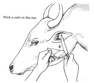
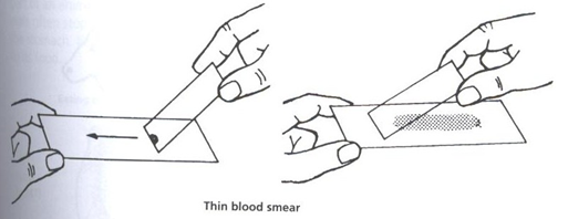
Blood smears can be taken from sick (or dead) cattle. In sick cattle prick the ear with a needle or the tip of a clean sharp knife and touch the drop of blood oozing out with a clean glass slide. With another slide touch the drop with one end of the second slide until the blood spreads along the angle between the two slides. Then push the upper slide along the lower slide so that it draws the blood after it. Wave the slide in the air until it is dry. Place inside a clean letter envelope and seal the envelope to prevent flies from getting in. Do NOT stick two smears together because it makes both useless for diagnostic purposes. Instead use one envelope per slide.
Also take a lymph gland smear from sick (or dead) cattle by inserting an 18 gauge needle attached to a syringe into a visibly swollen lymph node (best lymph node is the one in front of the shoulder blade), suck back on the syringe and expel the sucked up fluid onto a clean slide. Then let the slide dry and pack into a letter envelope for transport.
Blood samples are very useful for examining causes of diseases. Many diseases such as ECF, Babesiosis and anaplasmiósis are caused by microscopic disease organisms which will show up in a good blood or lymph node smear. Farmers can learn to make such blood smears and take them for analysis to the nearest vet or lab who has a microscope.
This will give a very accurate idea of the cause of the disease and will enable your vet to recommend the correct treatment. It is also much cheaper than tryng out different expensive drugs on the sick cow.
The procedure for making blood smears is simple:
-
Disinfect the inside of the ear with Dettol or Spiritus on a piece of cotton wool as well as the sharp knife to be used
-
Make a small prick in one of the blood vessels with a sharp pointed knife to draw just one drop of blood
-
Make sure the ONLY one drop of blood hits the middle of a clean glass slide and quickly draw a second glass slide through it to spread as thinly as possible on the lower slide. Wave the blood slide in the air for quick drying and place a clean glass slide on top of it to protect it from damage. Take the blood sample to the nearest vet office with a microscope.
 |
| Prick a vein in the ear to get a blood sample |
|
(c) William Ayako, KARI Naivasha
|
 |
| Thin blood smear |
|
(c) William Ayako, KARI Naivasha
|
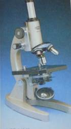 |
| A microscope for analyzing blood smears and other tissue samples in glass slides |
|
(c) William Ayako, KARI Naivasha
|
Glass slides are available from pharmacies and from some agro vet shops.
List of Kenya Government veterinary investigation laboratories under the ministry of livestock development and their contact addresses
|
No. |
Name of Laboratory |
Address |
Telephone Number |
Location |
Functions |
|
1 |
Veterinary Laboratory- Kabete
|
Private bag code 625 Kangemi Nairobi Kenya |
|
Kabete |
-Diagnosis of disease and parasites -Analysis of samples
|
|
2 |
Veterinary Investigation Laboratory |
P.O.Box Kericho Kenya |
|
Kericho |
-Diagnosis of disease -Analysis of samples
|
|
3 |
Veterinary Investigation Laboratory -Karatina |
P.O.Box Karatina Kenya |
|
Karatina |
Diagnosis of disease -Analysis of samples
|
|
4 |
Veterinary Investigation Laboratory-Nakuru |
P.O.Box 114 –Nakuru Kenya |
|
Nakuru |
-Diagnosis of disease -Analysis of samples
|
|
5 |
Veterinary Investigation Laboratory- Mariakani |
P.O.Box Mariakani Kenya |
|
Mombasa |
Diagnosis of disease -Analysis of samples
|
|
6 |
Veterinary Investigation Laboratory-Eldoret |
P.O.Box Eldoret Kenya |
|
Eldoret |
-Diagnosis of disease -Analysis of samples
|
|
7. |
Kenya Veterinary Vaccines Production Institute (KVVPI) |
P.O.Box 53280 Nairobi Kenya |
020-536043 /651595 |
Nairobi |
-Manufacture of veterinary vaccines |
Diagnostic disease chart for cattle in East Africa
Download here the Diagnostic chart on Cattle in pdf version
Lead symptom: Died suddenly – animal(s) not seen sick before death
All the diseases below can cause sudden death in cattle. The additional observations listed intend to guide you towards the most likely causes - but do not allow for confirmation of any particular disease. To get more information please follow the links below.
Additional observations:
| Un-clotted blood oozing from body openings (nose, anus), grazing in dry flood zone | -> Anthrax | |
| Swelling of muscle surface & gas under the skin (crackling sound), esp. 1 to 3 year olds | -> Blackquarter | |
| Feeding on clover / some legumes /green sorghum, abdomen extremely enlarged, froth in nose | -> Bloat | |
| Painful swelling on neck /brisket, extremely fast breathing, froth in nose | -> Haemorrhagic Septicaemia | |
|
Animals grazing wet area or flood-zone, in swamps and marshes |
-> Black Disease (Liver Fluke) | |
| Many ticks, only exotic cattle (= European breed) affected, convulsions, froth in nostrils | -> Heartwater | |
Other possible reasons why cattle can die suddenly are:
| Cattle have access to improperly stored chemicals, use of insecticide spray on/near cattle | -> Poisoning |
| Small bite marks on the head or leg | -> Snake bites |
| Sudden death only affecting suckling calves | -> see Calf problems |
Lead symptom: Coughing and/or pus and watery fluid coming from the nose
All the diseases below can cause respiratory disease in cattle. The additional observations listed will guide you towards the most likely causes - but do not allow for confirmation of any particular disease. To get more information please follow links below.
Additional observations:
Acute
|
After climatic/transport stress or crowding/mixing of animals, many very sick at once |
-> Pasteurellosis |
| Young animals suddenly sick, most recover, some don’t and become very sick | -> Calf Pneumonia |
| Very fast breathing, swollen lymph glands, froth in nostrils / mouth | -> ECF |
Chronic
| Deep dry cough, shallow fast breathing, grunt when exhaling, progressive loss of condition | -> CBPP |
| Occasional low moist coughing, mostly single adult animal, progressive loss of condition | -> Tuberculosis |
| Dry cough, chronic disease, esp. young animals on cool and wet highland pastures | -> Lung worms |
Lead symptom: Diarrhoea - scouring
All the diseases below can produce diarrhoea in cattle. The additional observations listed will guide you towards the most likely causes - but do not allow for confirmation of any particular disease. To get more information please follow links below.
Additional observations:
| Rains, esp. young cattle, feeding normally, poor body condition, not growing |
-> Worms Stom. & Intest. |
|
Few calves dying, some without diarrhoea, necrotic ear tips in calves, sporadic abortion |
-> Salmonellosis |
| Only young suckling calves affected by and dying from diarrhoea | -> Calf Scour |
| Mainly 8 months to 2 years old, dull, lesions inside mouth | -> Bovine Virus Diarrhoea/Mucosal Disease |
| Chronic diarrhoea in an adult, progressively loosing condition, feeding normally | -> Johne’s Disease |
| Acute, diarrhoea, lactating cow affected, fever, off-feed, udder is hot and swollen | -> Coli mastitis |
Lead symptom: Loosing condition, ribs sticking out, rough coat
With all the diseases listed below cattle do severely loose condition. The additional observations listed will guide you towards the most likely causes - but do not allow for confirmation of any particular disease. To get more information please follow links below.
Additional observations:
| Feeding normally, rainy season, mainly young cattle, often diarrhoea | -> Worms Stomach & Intestine |
| Feeding normally, weak, anaemia, sometimes diarrhoea | -> (Blood-sucking) Worms Stomach & Intestine |
| Feeding normal, bottle jaw (= oedema on lower jaw), grazing in marsh / near swamp | -> Liver Fluke |
| Very sleepy, pale membranes (around eyes), large lymph glands, area has Tse-Tse flies | -> Trypanosoma |
| Feeding normally, dull or abnormally coloured hair coat | -> Mineral deficiency |
| Dry cough, ongoing respiratory disease problem in the herd since weeks and months | -> CBPP |
| Occasional low cough, sometimes also diarrhoea, mostly single adult animal | -> Tuberculosis |
| Chronic diarrhoea in an adult, progressively loosing condition, feeding normally | -> Johne’s Disease |
| Very thin, drought or insufficient access to quality feed; very hard coarse silage | -> Starvation |
| Wet, grazing inside thicket, very high tick load, pale membranes (around eyes) | -> Tick worry |
Lead symptom: Abortion
All the diseases below can cause abortions in cattle. The additional observations listed will guide you towards the most likely causes - but do not allow for confirmation of any particular disease. To get more information please follow links below.
Additional observations:
|
Most foetuses are expelled near term, very often retained placenta after the abortion |
-> Brucellosis |
| Very strong rains, females abort at all stages of pregnancy, newborns dying | -> Rift Valley Fever |
| Late abortion, fetus decomposed, animals fed on poor quality silage (bad smell) | -> Listeriosis |
| Late abortion, wet pasture (standing puddles), cow may be sick before abortion (jaundice) | -> Leptospira |
| Some calves dying without clear signs, some animals with severe diarrhoea | -> Salmonella |
| Abortion between 4 and 6 months of pregnancy (often unnoticed), birth of paralysed calf | -> Neospora |
| Early abortion (first 4 to 5 months hence often unnoticed), poor herd conception rate | -> Trichomonas |
Lead symptom: Pale membranes or jaundice, often swollen lymph glands
With the diseases listed below cattle can develop anaemia (visible as pale/white membranes around the eyes) or jaundice (yellow membranes around the eyes) and have swollen glands. The additional observations listed will guide you towards the most likely causes - but do not allow for confirmation of any particular disease. To get more information please follow links below.
Additional observations:
| Adult animal, first pale membranes later jaundice, dark-red urine, constipation/diarrhoea | ->Babesia |
| Esp. in adults, membranes first pale then yellow, constipation/diarrhoea-similar to Babesia | ->Anaplasma |
| Large glands, fever, cloudy eyes, fast breathing, sometimes diarrhoea, very sick & getting worse | -> ECF |
| Large glands, deteriorating slowly, very weak, always sleepy, pale membranes, area has Tse-Tse | ->Tryps |
| Normal feeding, anaemia, normal glands, sometimes diarrhoea | ->(Blood-sucking) Worms Stom. & Intestines. |
Lead symptom: Lesions on the skin
All the diseases listed below can produce lesions on the skin of cattle. The additional observations listed will guide you towards the most likely causes - but do not allow for confirmation of any particular disease. To get more information please follow links below.
Additional observations:
| Painful nodules on skin (later become small wounds), inflamed eyes, running nose | -> Lumpy Skin |
| Tufts of hair coming off easily (esp. neck & shoulder), bold plaques, scabs, during rains | -> Dermatophilus |
| Round hairless skin lesions, become confluent, not itchy, esp. in young animals | -> Ringworm |
| Very itchy skin lesion with hair loss and thickening of skin (can look like elephant skin) | -> Mange |
| Affects light coloured skin parts most exposed to sun, skin sloughing off, not itchy | -> Photosensitisation |
| Rubbing, scratching and biting the skin, sometimes also anaemia | -> Lice (massive infection) |
| Bleeding spot on skin, attracts flies and does not heal for a long time | -> Filaria |
Lead symptom: Lesions in the mouth and on the head
All the diseases listed below can produce lesions inside the mouth, on the head and/or affect the eyes of cattle. The additional observations listed will guide you towards the most likely causes - but do not allow for confirmation of any particular disease. To get more information please follow links below.
Additional observations:
| Drooling, lameness, erosions on tongue / gums and also between the claws | -> Foot & Mouth Disease |
| Very sick(!), inflamed eyes (milky-blue), ulcers in mouth, crusts on nostrils | -> Malignant Catarrhal Fever |
| Weeping, one eye (rarely both) affected, eye: cloudy/white/blue/milky, temporary blind | -> Pink Eye |
| Small blood spots in the eye - come and go (also inside vulva), nose bleeding, ‘poached egg eye’ | -> Ondiri |
| Lesions inside mouth, weeping, mainly 8 months to 2 years old, severe diarrhoea | -> Mucosal Disease |
| Weeping (both eyes), eyes cloudy, very sleepy, poor condition, large lymph glands, Tse-Tse flies | -> Tryps |
| Very sick, cloudy / milky eyes, high fever, fast breathing, swollen lymph glands | -> ECF |
| Bottle jaw, grazing in or near marsh/swamp | -> Liver fluke |
Lead symptom: Paralysis / Central nervous disorder / circling
With all the diseases listed below cattle can be paralysed or show abnormal movement and behaviour. The additional observations listed will guide you towards the most likely causes - but do not allow for confirmation of any particular disease. To get more information please follow links below.
Additional observations:
|
Single animal affected, drooling, knuckling of hind fetlock, trying to pass faeces, bellowing |
-> Rabies |
| Coordination problems, exotic cattle affected, convulsions, froth in nostrils before death | -> Heartwater |
| Fed on poor silage, drooping ear, blind, circling, tongue hanging out, also abortions | -> Listeriosis |
| Stiff, unable to stand up, can’t swallow, flaccid tongue, slight bloat | -> 3-Day Sickness (Ephemeral Fever) |
| Very good milker in peak lactation, sleepy, sometimes licking & biting itself, can get excited | -> Ketosis |
| Older cow immediately after calving, too weak to stand up but trying | -> Hypocalcaemia (milk-fever) |
| Blindness, head pushing, aimless wandering, slowly progressing, become recumbent | ->Tapeworm (cyst) |
Lead symptom: Lameness
With all the diseases listed below cattle can go lame. The additional observations listed will guide you towards the most likely causes - but do not allow for confirmation of any particular disease. To get more information please follow links below.
Additional observations:
Common causes of lameness in cattle are foot problems due to injury and lack of foot care:
| -> Abscess of the heal, the coronary band, the white line, the sole |
| -> Foreign body (stone) between the claws or penetrating the sole |
| -> Ulcers of the sole |
| -> Footrot (cheesy material in lesion between the claws, very smelly) |
| -> Crack of the hoof wall |
| -> Overgrown claws (corkscrew claw, scissor claw) |
Infectious causes of lameness in cattle:
| Drooling, erosions on tongue & gums and also between the claws | -> Foot & Mouth Disease |
| Some unable to stand up, can’t swallow, flaccid tongue, slight bloat | -> 3-Day Sickness (Ephemeral Fever) |
| Swelling of muscle, gas under the skin (crackling sound), esp. 1 to 3 year olds | -> Blackleg/Blackquarter |
| Drooling, knuckling of hind fetlock, trying to pass faeces, bellowing | -> Rabies |
Diagnostic disease chart for sheep and goats in East Africa
Download here the Diagnostic chart on Sheep & Goats in pdf version
Lead symptom: Died suddenly – animal(s) not seen sick before death
All the diseases below can result in sudden death of sheep & goats. The additional observations listed intend to guide you towards the most likely causes - but do not allow for confirmation of any particular disease. To get more information please follow links below.
Additional observations:
| Esp. young sheep affected, grazing on fresh pasture - short grass | -> Enterotoxaemia |
| Un-clotted blood oozing out, grazing in dry flood zone | -> Anthrax |
| Bloat, uncontrolled access to grain (maize or other) | -> Acidosis |
| Wet conditions, many ticks, often exotic sheep/goats (= European breed) | -> Heartwater |
| Esp. young sheep affected, abrupt change in feed | -> Septicaemic Pasteurellosis |
| Esp. young sheep in flood zone/swamp, also anaemia & bottle jaw | -> Black Disease/Liver fluke |
| Grazing after/during rain on/near recently fertilized field | -> Nitrite Poisoning |
| Only suckling lambs and kids affected | -> Lambs and kids problems |
Lead symptom: Coughing and/or pus coming from the nose
All the diseases below can produce respiratory signs in sheep & goats. The additional observations listed will guide you towards the most likely causes - but do not allow for confirmation of any particular disease. To get more information please follow links below.
Additional observations:
| Only goats affected and dying, no sheep with respiratory signs | -> CCPP |
| Climatic stress, all age groups of affected, mostly young animals dying | -> Pneumonic Pasteurellosis |
| Pox lesions on head and skin, lambs & kids with respiratory disease signs | -> Sheep and Goat Pox |
| Many sheep & goats affected, also diarrhoea, most adult goats dying! | -> PPR |
| Feeding normally, pus in the nose, sneezing especially in the morning | -> Nasal Bot Fly |
| Feeding normally, chronic cough, animals kept in cool highland region | -> Lung Worms |
| Only young animals affected | -> Pneumonia of the young |
Lead symptom: Diarrhoea - scouring
All the diseases below can produce diarrhoea in sheep & goats. The additional observations listed will guide you towards the most likely causes - but do not allow for confirmation of any particular disease. To get more information please follow links below.
Additional observations:
| Rainy season, feeding normally, not putting on weight | ->Round-Worms |
| Also respiratory signs, most adult goats dying! | ->PPR |
| Newborn lambs/kids all dying, very strong rains, mass abortions | ->Rift Valley Fever |
| Many sheep (incl. adults) dying from acute diarrhoea, some abortions | ->Salmonellosis |
| Small number affected, severe respiratory signs, sheep introduced recently | ->Nairobi Sheep Disease |
| Only lambs and kids affected | ->Diarrhoea of the young |
| Only in lambs/kids, sporadic colic, no serious scouring, flat worms (“spaghetti”) in faeces | ->Tapeworms |
Lead symptom: Loosing condition and showing a rough coat
With all the diseases listed below sheep & goats can be in very poor condition. The additional observations listed will guide you towards the most likely causes - but do not allow for confirmation of any particular disease. To get more information please follow links below.
Additional observations:
| Feeding normally, not dewormed for long, often diarrhoea | -> Round Worms |
| Pale around eyes, weak, feeding normally, sometimes diarrhoea | -> Blood-sucking Round Worms |
| Bottle jaw, pale eye membranes, feeding normally, grazing in marsh / near swamp | -> Liver Fluke |
| Sleepy, pale membranes (around eyes), kept in humid warm lowlands with Tse-Tse flies | -> Trypanosoma |
| Feeding normally, dull or abnormally coloured hair coat (pale rings around eyes) | -> Mineral deficiency |
| Feeding normally, dull or abnormally coloured hair coat, lambs/kids with swayback | -> Copper deficiency |
| Ongoing severe respiratory disease problem in goat herd since weeks | -> CCPP |
| Very thin, drought, animals only have access to very poor quality feed | -> Starvation |
| Wet, grazing inside thicket, very high tick load | -> Tick worry |
| Animals irritated, rubbing/kicking the skin, poor condition, pale eye membranes | -> Lice (mass infestation) |
| Animals itching and rubbing their skin, loosing condition, bold skin with dry scabs | -> Mange |
Lead symptom: Abortion
All the diseases below can cause abortions in sheep & goats. The additional observations listed will guide you towards the most likely causes - but do not allow for confirmation of any particular disease. To get more information please follow links below.
Additional observations:
| Many abortions, most foetuses near term, often retained placenta after the abortion | -> Brucellosis |
| Fresh dead lambs & kids expelled almost on term | -> Enzootic Abortion of Ewes |
| Sporadic abortions, often with retained placenta, poor herd fertility | -> Q-Fever |
| Very strong rains, almost all pregnant females abort, newborns dying | -> Rift Valley Fever |
| Animals were fed silage | -> Listeriosis |
| Some sheep with severe diarrhoea, lambs dying | -> Salmonella |
| Sporadic abortions, dark urine, jaundice around the eyes | -> Leptospirosis |
Lead symptom: Lameness & Stiffness
With all the diseases listed below sheep & goats can become stiff or go lame. The additional observations listed will guide you towards the most likely causes - but do not allow for confirmation of any particular disease. To get more information please follow links below.
Additional observations:
| Many affected, feeding normally, loosing condition, smelly lesions on feet | -> Foot Rot |
| Many affected, fever, salivation | -> Foot & Mouth Disease (FMD) |
| Few affected, small abscess between the claws | -> Untreated injury (thorns) |
| Many affected and going stiff, swollen face, fever, salivation, crusts around nose | -> Blue Tongue |
| Stiffness, tremors, spasms, quickly getting worse, stiff tail, animal stands like a sawbuck | -> Tetanus |
Lead symptom: Lesions and swellings of the skin
All the diseases listed below can produce lesions and swellings on the skin in sheep & goats. The additional observations listed will guide you towards the most likely causes - but do not allow for confirmation of any particular disease. To get more information please follow links below.
Additional observations:
| Pox lesions, scabs and crusts on lips, mouth and on the head, only in the young | -> Orf |
| Pox lesions, scabs/crusts on lips head and on whole skin, pneumonia in young | -> Sheep & Goat Pox |
| Open wound with pus, swelling oozing pus, hot painful swelling | -> Wounds and Abscesses |
| Swelling oozing smelly liquid or pus, contains small larva, attracts flies, sheep/goat very sick | -> Fly Strike |
| Animals in poor condition, itching and rubbing, loosing hair, bold skin, loose crusts | -> Mange |
| Itching / rubbing, loosing hair, bold/rough skin, loose crusts ONLY on limbs and scrotum | -> Foot Mange |
| One or two lymph-glands hard and swollen, contain dry or creamy pus | -> Pseudotuberculosis |
| Round hairless skin lesions with crusting, sheep/goat housed together with cattle | -> Ringworm |
Lead symptom: Lesions inside the mouth and on the head
All the diseases listed below can produce lesions inside the mouth, on the lips or on the skin of the head in sheep & goats. The additional observations listed will guide you towards the most likely causes - but do not allow for confirmation of any particular disease. To get more information please follow links below.
Additional observations:
| Pox lesions, scabs and crusts on lips, mouth and head, only in the young | -> Orf |
| Pox lesions, scabs/crusts on lips head and on skin, pneumonia in the young | -> Sheep & Goat Pox |
| Lameness, fever, salivation | -> Foot & Mouth Disease (FMD) |
| Stiffness / lameness, swollen face, salivation, crusts around nose and eyes | -> Bluetongue |
Lead symptom: Paralysis / abnormal movement & behaviour / circling
With all the diseases listed below sheep & goats can be paralysed or show abnormal movement and behaviour. The additional observations listed will guide you towards the most likely causes - but do not allow for confirmation of any particular disease. To get more information please follow links below.
Additional observations:
| Mostly single animal affected, circling movement | -> Hydatid cysts |
| Mostly single animal affected, paralysis (most common sign!), also hypersexual behaviour | -> Rabies |
| Group affected, coordination problems, circling movement, paddling on side before death | -> Heartwater |
| Group affected, fed on silage, tilted head, drooping ear, circling movement, abortions | -> Listeriosis |
| Lambs/kids show trembling twitching and swayback, adults with pale/dull hair | ->Copper deficiency |
| Stiffness, tremors, spasms, quickly getting worse, stiff tail, animal stands like a sawbuck | -> Tetanus |
Review Process
- Author September 2011: Dr Hugh Cran, Veterinary Practitioner, Nakuru Kenya
- Review May 2013: Dr Mario Younan (DVM, PhD), Regional Technical Advisor for VSF-Germany, working in East Africa since 1995
Information Source Links
- John Gilmour 1992: Making the Most of Ovine Necropsy. In Practice Journal of Veterinary Postgraduate Clinical Study, Volume 14, No 3, May. ISSN No 0263/841 X
- MJ Fitzpatrick 1950: Notes on Animal Diseases, The Diagnosis of Animal Diseases in the Veterinary Laboratory. Veterinary Research Laboratories, Veterinary Department Kabete, Kenya. Originally printed in the East African Agricultural Journal.
- Blood, D-C., Radostits, O.M. and Henderson, J.A. (1983) Veterinary Medicine - A textbook of the Diseases of Cattle, Sheep, Goats and Horses. Sixth Edition, Bailliere Tindall London. ISBN: 0702012866
