Haemorrhagic Septicaemia
Local names:
Borana: quando / Samburu: nalngiarrngarri / Somalia: kharar / Turkana: angaare
Description: Respiratory disease
Introduction
Haemorrhagic Septicaemia also sometimes called "shipping fever" is an acute bacterial disease of cattle and water buffaloes and pigs. It is caused by Pasteurella multocida serotypes B2 in Asia and E2 in Africa. Also Fowl Cholera is caused by Pasteurella sp.
Outbreaks of acute pasteurellosis caused by other serotypes of P. multocida or by P. haemolytica should not be called Haemorrhagic Septicaemia, even though septicaemia may be a feature.
The disease is endemic (insert dictionary) in south and east Asia. Occasionally outbreaks occur in Africa, southern Europe and the Near East.
Mode of spread
Only water buffalo and cattle are regularly affected. Stress factors, coupled perhaps with viral or other infections, weaken the animals' defences and predispose them to the disease. The disease in Africa may suddenly appear and then may not reappear for many years. The worst outbreaks occur during the rainy season.
- Transporting and herding animals may trigger an outbreak.
- Spread within a group usually occurs at night when animals are enclosed, and between groups via communal watering points.
- In areas where the disease is prevalent, small numbers of healthy animals - cattle and water buffalo - carry the bacteria in the nasopharynx (the area of the upper throat that lies behind the nose) or tonsils and act as reservoirs of infection. Bacteria are then spread through the air.
- During an outbreak sick animals excrete large numbers of bacteria in runny noses, saliva and faeces.
- For several weeks after an outbreak 20 - 50% of animals carry the organism and are capable of infecting susceptible in-contact animals.
- Indirect transmission can also occur.
- The causal bacteria can survive for several hours in moist conditions, but die rapidly if exposed to sunlight or thorough drying
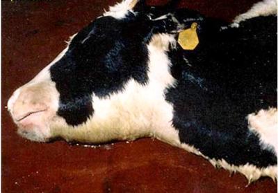 |
| Signs of Haemorrhagic Septicaemia |
|
© USDA
|
Signs of Haemorrhagic Septicaemia
In areas where the disease is prevalent, animals over 1 year old have often acquired immunity from previous outbreaks, therefore disease is more common in young stock. Outside such areas, clinical cases are seen in all age groups.
- Occurrence rates range from low to high, but in general death rates are over 50% and may approach 100%.
- Most cases are acute or peracute, resulting in death within 8 - 24 hours after onset. Because the course is so short, clinical signs may easily be overlooked.
- The infection is thought to first increase in the region of the tonsils. In susceptible animals, blood poisoning develops rapidly, and death due to the presence of toxins produced by the bacteria follows within 8 - 24 hours after the first signs develop.
- The first signs are dullness, reluctance to move and fever.
- Excess salivation and a clear nasal discharge appear.
- A common feature is oedema (swelling) of the laryngeal (throat) region, which spreads down to the brisket, and up to the region of the parotid gland near the ear. It sometimes involves the whole head.
- The tongue may swell and protrude and mucous membranes are congested. Affected animals have difficulty in breathing.
- Most animals collapse and die within a few hours. Recovery is rare. Occasionally an animal may remain sick for several days. Death is due to suffocation and to the release of toxins produced by the bacteria.
Diagnosis
Rapid course of illness, high herd incidence, weather conditions, fever, typical throat swellings, coupled with the characteristic post mortem findings, are suggestive of Haemorrhagic Septicaemia.
Anthrax, gas gangrene caused by clostridial bacteria, certain snake bites, and pneumonic pasteurellosis should be considered, as these can occasionally give rise to similar swellings.
- Post-mortem reveals extensive oedema of the head, throat and brisket. The fluid is straw coloured and infiltration may extend from the subcutaneous tissue into the muscle.
- Numerous small haemorrhages are found throughout the carcass.
- Blood-tinged fluid may be found within the pericardial sac and in the chest and abdominal cavities.
- The pharyngeal and cervical lymph nodes are swollen and frequently contain small haemorrhages.
- In those animals which have survived for several days pneumonia with thickening of the interlobular septae may be seen, but well- established bronchopneumonia is more likely to be caused by other serotypes of P. multocida or P. haemolytica than in true Haemorrhagic Septicaemia.
- When calves are affected haemorrhagic gastroenteritis may be seen.
To confirm the diagnosis, a veterinary surgeon will take samples of tissue from blood, lung, liver and spleen and send these to a laboratory to determine the organism that has caused the disease. If the animal has been dead for longer than 8 hours, the vet will send a long bone.
Diseases with similar symptoms
- Swollen lymph nodes, fever and froth discharge from the mouth: See ECF and MCF (under construction)
- Anthrax, Salmonellosis, Rinderpest - see below
Prevention - Control - Treatment
Treatment
To be of value, treatment must be started early; but because of the rapid course of the disease, this is often not practicable. Treatment is of little value once animals appear shocked, and may instead cause a deadly crisis by causing death of organisms and the release of toxins produced by the bacteria.
Various sulphonamides, tetracylclines, penicillin and chloramphenicol are effective if administered in the very early stages.
Prevention
- The principal means of prevention is by vaccination, which in areas where the disease is prevalent, should be carried out on an annual basis.
- Where the disease is not prevalent, the cost of regular vaccination may not be justified, but when an outbreak occurs, vaccination should be considered in order to control the spread of infection.
- Herd quarantine, the segregation of sick animals from healthy ones, and routine disinfection all help to slow the spread of disease between and within herds.
- In previously infected herds, natural immunity exists and occurrence rates and death rates are substantially lower than in herds not previously infected. Immune animals, however, can carry and transmit infection.